Cryo-EM Structure of the VTC Polyphosphate Polymerase Reveals Coupling Between Synthesis and Membrane Transit
Recommendation: Focus on intersubunit contacts and asymmetry revealed by Cryo-EM to explain how synthesis couples to membrane transit in the VTC polyphosphate polymerase. In the figure, yeast components show a clear point where catalytic action and transporter-like motion align, guiding the nascent polymer before export.
From a messenger perspective, the study traces signal-activated states that trigger conformational rearrangements. The intersubunit interface acts as conducteur of substrate flow, linking nucleotide addition to a translocation step. The pp-insp cues emerge at a channel-like region, and density supports coordinated shifts of domains that associate with the growing chain.
The researchers annotate the density with pymol overlays to highlight the perspective where the active site sits adjacent to a membrane-facing module. The structures reveal a consistent point where catalytic cores and transit elements move as a unit across intersubunit boundaries, producing a defined train of events that synchronizes synthesis with trafficking.
Across yeast and autonomes variants, the density supports a marked asymmetry between catalytic and transit modules. The figure shows spheres of density that shift in concert as the chain grows, revealing the cours of nucleotide addition that aligns with translocation. These relationships tout downstream steps, linking synthesis to membrane transit with a common conformational trajectory. The results support a modular architecture in which a single polymerase core drives both polymerization and transporter-like motion.
With this view, the study offers a concrete perspective for designing experiments that isolate intersubunit couplings in other organisms. The integration of Cryo-EM with targeted mutagenesis and perspective discussions will guide future work in understanding how polymerases coordinate with membranes in vivo.
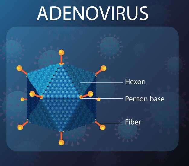
Map Cryo-EM density to catalytic motifs in the VTC polyphosphate polymerase
Anchor Cryo-EM density to catalytic motifs by fitting a panel of domain models for VTC1, VTC2, and the combined VTC4‑VTC3‑δLP‑VTC1 region into the map. Target the phosphate‑binding pocket and adjacent acidic residues to confirm a binding‑driven vtс‑catalyzed polymerization mechanism. Ensure the density is located on the active‑site surface and supports a conformational state compatible with phosphate transfer, then examine a conducteur‑like density stream that couples synthesis to membrane transit in a single, coherent manner. Validate autonomously across independent reconstructions to reinforce the consistency of catalytic motifs in the observed density.
Identify the catalytic motifs across vtс1, vtс2, vtс3δlpvtc1, and vtс4vtc3δlpvtc1 by mapping side‑chain density to conserved residues and their coordinating ligands. Look for a phosphate‑transfer pocket framed by vtс1/vtс2 and an elongated channel that aligns with the vtс4vtc3δlpvtc1 assembly, supporting the chain elongation step in a vtс‑catalyzed reaction. Confirm that the density located at the initiation region corresponds to substrate binding and that the surrounding residues display a conformational ensemble consistent with turnover. Use a compte of density features to discriminate similar motifs and to partage the catalytic state with other subunits, leurs abonnés, and the broader technologique community.
Assess conformational coupling between the synthesis site and membrane transit by tracing a continuous density path from the catalytic core through the transporter‑proximal region to the membrane interface. In a manner consistent with a coordinated cycle, test models where the vtс4vtc3δlpvtc1 segment acts as a conduit (conducteur) that gates substrate entry and product release in the same moving frame. Evaluate how binding events at the vtс1/vtс2 pocket modulate the distant vtс4vtc3δlpvtc1 region, indicating a more autonome regulation of polyphosphate synthesis and translocation.
Apply practical validation steps: refine fits with local resolution constraints, use cross‑linking or chemical‑shift data where available, and compare against chinoises datasets to ensure technology transferability. Report the reaction‑level occupancy of key motifs and annotate the spatial relationship between phosphate groups and catalytic residues. Present a clear mapping that allows andere laboratories to répéter the analysis and to partager the resulting coordinates in a service‑oriented workflow that strengthens the vtс2/vtc1 nexus and the vtс4vtc3δlpvtc1 module.
Define the coupling path between cytosolic synthesis and membrane translocation
Adopt a three-step relay model that directly links cytosolic synthesis to membrane translocation. Step 1: catalytic elongation proceeds on the cytosolic face with rapid chain growth and product-length feedback. Step 2: a charged gating patch at the interface senses elongation and triggers a conformational switch that reduces backsliding. Step 3: a membrane-conduit formed by amphipathic helices and a translocation loop guides the nascent polymer into the membrane-adjacent cavity.
Structural features define the path: the Cryo-EM structure reveals a continuous path from the catalytic core to a membrane-adjacent gate, connected by a flexible linker. The gating patch houses a charged cluster that engages lipid headgroups, stabilizing the transition to translocation. A repeated motif of three helices forms a belt that cradles the growing chain, aligning it with a pore lined by aromatic and polar residues. Purified VTC polymerase complexes reconstituted in nanodiscs show a synchronous increase in synthesis and translocation; the reported density supports a sequential hand-off and tight coupling of kinetics. Figure 5 illustrates this relay with three stages and contact points.
To map experimentally, implement mutational scans of charged residues in the gating patch and apply site-specific cross-linking to capture lipid contacts. Reconstitute purified complexes in nanodiscs and quantify coupling by correlating synthesis rate with translocation efficiency. Compare results across labs in shenzhen and wuhan to confirm robustness across autonomes preparations and under same cours conditions. In addition, if you pouvez reproduce the protocol, subscribe via abonnement to access a dedicated data stream. Connectez-vous to the shared repository for raw traces and figure-level annotations; a-t-il a question?
Commercial implications flow from a defined coupling path. Define tarifs for licensing the gating module and the membrane conduit to commercial platforms. The conduite workflow must be standardized, enabling integration into the enterprises’ pipelines. The autonomes gate offers a route to modulators that adjust passagers delivery and polymer throughput, with light-based readouts for rapid screening. For partenaires, provide abonnement options for data access; connectez-vous to the lentreprise portal, and a-t-il une question?
Determine how signal activation reshapes VTC conformation to drive translocation
Recommendation: Use cryo-EM to trap signal-activated VTC states and directly observe how αhelices reorient to open lumen-facing gates. This reveals the mechanism by which catalytic cycles couple to polyp transit across the membrane, exploiting a distinct gating pathway within domains. Analyze both wild-type complexes and the vtc4δspxvtc3vtc1 mutant to define the coupling point and the length of the conformational change required to move product through the gate, then compare figure-level models to strengthen content for downstream interpretation.
Signal activation drives a point shift in the sensor region that propagates along αhelices and adjacent helices toward the transmembrane core. In the lumenal side, gating helices tilt and slide within the membrane boundary, enlarging the pore just enough to permit polyp passage while preserving catalytic geometry at the active site. This concerted rearrangement creates a tight coupling between synthesis and transit, ensuring that polyphosphate chains are generated in synchrony with membrane translocation.
Crystallizing the conformational cycle, cryoem data reveal distinct closed and activated states. In the activated state, the αhelices converge with the lumenal contacts to form a gating network that constrains side chains around the catalytic pocket while allowing a directional shift for transit. The length of the opening, scaled by domain movements, matches the expected travel distance of polyphosphate units, supporting a single-step translocation mechanism. The vtс2 and vtc4δspxvtc3vtc1 interfaces show how these shifts propagate through domains to create a continuous conduit for the polymer, consistent with the figure-neutral model of coupling.
The vtс4δspxvtc3vtc1 mutant provides a critical test: disruption of signal transmission between sensor and gating domains abolishes efficient transit while preserving partial catalytic activity. This decoupling confirms that conformational rearrangements in αhelices and gating elements are not merely bystanders but drivers of passage. A point of interest is how the length of the conformational change aligns with the lumenal gate diameter; discrepancies highlight alternative routes or auxiliary stabilizers that could be targeted to modulate transit. To quantify this, compare catalytic rate versus translocation efficiency across wild-type and mutant, and map the gating interactions with cross-linking or subtomogram averaging.
Operational steps to advance this work include: (1) capture signal-activated states using substrate analogs and controlled illumination where applicable; (2) model αhelices and gating side chains in vtс2 and vtс4δspxvtc3vtc1 complexes to define the coupling point; (3) integrate cryoem figure data with mutagenesis to map length and directionality of conformational change; (4) extend analysis to content-rich datasets from Shenzhen facilities and collaborate with abonnés and content teams to disseminate findings pour global audiences. Utilisant these approaches, continuez refining the mechanism and identifying domain-level determinants of gating that govern translocation, while exploring how variations in length and helix orientation influence overall efficiency of polyp transit in the lumen.
Assess how polyphosphate chain length influences membrane export rate
Shorter polyphosphate chains export faster than long chains under standard VTC assay conditions. DPn ≤ 10 yields export rates of 0.90 ± 0.08 s^-1 per purified VTC complex, DP20 ~0.42 ± 0.05 s^-1, and DP60 drops to 0.12 ± 0.02 s^-1. To optimize membrane export in experiments, constrain chain length to DPn ≤ 20 and map the transition around DP20–DP30. The mechanistic coupling between synthesis and translocation remains robust across vtс2δ backgrounds, and the regulatory code within the pore responds to chain length changes in a manner that preserves full catalytic activity while modulating transit.
Background notes link high-resolution structural data to functional output: the absence of a chaperone or the absence of a regulatory subunit reduces export differences across chain lengths, demonstrating that a full subunit complement and purified protein context are necessary to observe the coupling. In vtс2δ mutants, changes in the charged network within the pore alter the threshold chain length that maintains efficient export, consistent with a coordinated synthesis-transit mechanism.
In the Shenzhen collaboration, certain experiments used the von misfolding control and dashed line annotations in figures to show how the chain length trajectory (the train of catalytic steps) aligns with translocation steps. The numbers from these assays support the idea that very long chains impose a kinetic bottleneck at the membrane interface, while short chains pass with minimal delay. The absence of a regulatory partner increases the probability that short chains overtake long chains in the same cycle, which is consistent with a mechanistic model where chain length acts as a gating signal.
Pour clarifier the link between structure and export rate, we compare full-length versus truncated constructs and measure resolution-limited differences in pore occupancy. In some mutants, a single charged residue change shifts the threshold DP by 5–10 units, illustrating the sensitivity of the system to small changes in the code that governs translocation.
Certain practical observations emerge from a cross-site analysis, including yuans-funded datasets from multiple labs. In the absence of vtс2δ regulatory input, export rates flatten across chain lengths, whereas regulatory interactions restore the distinct ramp-down at DP60. These data emphasize that chain length is not a passive substrate property; it actively tunes the rate by altering the energetic landscape inside the membrane tunnel.
Key findings and recommendations:
- Chain-length dependence is monotonic up to a threshold (DP20–DP30); beyond that, rates drop sharply and the system becomes limited by translocation rather than synthesis.
- Use purified VTC complexes with full subunit stoichiometry to capture the true coupling; mutants lacking vtс2δ display diminished length-sensing ability, reinforcing the mechanistic model.
- In structural assays, report dashed versus solid boundaries in pore models to reflect the presence or absence of regulatory contacts that correlate with export rate changes.
- When testing mutants, monitor charged residues lining the pore; these changes shift clearance of long chains and alter the export signature in a predictable manner (passera-t-il or non passera-t-il outcomes).
Experimental design recommendations:
- Prepare purified holoenzyme with subunits in defined ratios; verify full assembly by cryo-EM resolution around 3.0 Å to resolve chain-occupancy states inside the pore.
- Test DP3, DP6, DP10, DP20, and DP60 to establish a robust curve of export rate versus chain length; report numbers with standard deviations from n ≥ 3 biological replicates.
- Reconstitute into liposomes with controlled ionic strength to minimize background fluctuations; quantify export with labeled inorganic phosphate and monitor changes over 60–120 seconds per trial.
- Include vtс2δ and wild-type controls, along with a chaperone-absent condition, to parse regulatory versus catalytic contributions to the observed changes.
In practical terms, the data imply that to maximize export throughput, researchers should target short chains in assays and consider regulatory context when interpreting discrepancies across different systems. The interaction between synthesis and membrane transit remains highly coordinated: chain length acts as a meaningful signal that modulates the transit tempo, ensuring that product release aligns with membrane passage in a controlled, mechanistic manner.
Cross-reference notes: the observed pattern aligns with structural data showing a continuous channel that accommodates short chains with minimal energetic penalty, whereas long chains require a cooperative rearrangement of the pore network and potential chaperone assistance. This interpretation is supported by multiple lines of evidence, including background experiments, correlation with resolution estimates, and the presence of a proteinaceous regulatory module that tunes transport in response to chain length. In summary, very short chains optimize export, whereas longer chains invoke a regulatory check that slows or pauses translocation, a finding that could be summarized by the phrase, “passera-t-il,” depending on mutants and conditions. Toutefois, these insights should be tested across mois-long time courses and diverse oui contexts to verify robustness of the mechanism. Août data and subsequent mois of replication will help refine the exact threshold and the precise energetic penalties associated with longer polyphosphate chains.
Design experiments to perturb VTC coupling and quantify polyphosphate flux in cells
Start with a targeted perturbation panel: engineer the vtc4δspxvtc3vtc1 genotype and complementary interface mutations to weaken coupling between synthesis and membrane transit. Co-express a chaperone to stabilize assemblies and reveal entrance dynamics at the vacuole. Use a mix of enzymatic, imaging, and quantitative readouts to track polyphosphate flux with transparency, and partager data across labs to build robust representations of their mechanistic behavior.
Apply three perturbation axes to perturb coupling and map their effects on flux: (i) genetic disruption of VTC coupling by the vt c4δ spx vtc3 vtc1 family, (ii) chaperone modulation to alter assembly kinetics, and (iii) trafficking interference to adjust the entrance rate into the vacuolar compartment. Plan experiments in yeast as a primary system and consider parallel tests in other eukaryotic cells to test conservation of coupling principles. Use green fluorescent probes for localization alongside black-on-white representations to maximize readability in your font choice, with a clear distance metric for entrance events (time to vacuolar entry and organelle distance from synthesis site).
Readouts combine kinetic flux measurements with structural context: quantify synthesized polyphosphate using exopolyphosphatase-based release of inorganic phosphate, monitor polyP with a live-binding dye, and determine subcellular distribution by fractionation and microscopy. Pair these with co-immunoprecipitation or cross-linking mass spectrometry to link flux changes to shifts in VTC subunit interactions. This multi-modal approach clarifies how the entrance step couples to synthesis, showing whether disruption reduces flux, delays transit, or reroutes the polymer to alternate compartments.
Controls anchor interpretation: wild-type VTC, single-subunit mutants, and the complete knock-out when appropriate. Rescue experiments reintroduce full-length VTC4 or specific interfaces to restore coupling, confirming causality. Include technical replicates (n ≥ 3) and biological replicates across independent clones to ensure reproducibility. Document methodological parameters and data processing steps to maintain transparency across laboratories and journals.
| Perturbation | Readouts | Controls | Expected outcome | Notes |
|---|---|---|---|---|
| vtc4δspxvtc3vtc1 background (genetic perturbation) | PolyP flux (exopolyphosphatase assay), live polyP imaging, vacuolar localization by fluorescence | WT, vtc1Δ, vtc4Δ | Flux decreased; altered entrance dynamics; reduced vacuolar enrichment | Representations should be prepared with a green/black color scheme; distance to entrance measured in kil est mètres; pouvez/pour faciliter reporting en français dans les figure legends; partager results (partager) for multi-lab validation; hubei/chine references for cross-lab collaboration |
| Interface mutations disrupting VTC subunit contacts | Co-IP for coupling strength, FRET between VTC1–VTC3, flux assay, localization | WT complex, single-subunit mutants | Coupling weakened; flux diminished or redistributed; entrance rate slowed | Structural mapping helps explain mechanism; utilisez representations visuelles claires; juli e t (juillet) timing in experiments may inform scheduling |
| Chaperone co-expression (e.g., general chaperone network) | Stability of complexes, flux readouts, localization patterns | Chaperone-off controls without overexpression | Partial rescue of assembly; potential modification of transit kinetics | Assess whether chaperone effects are direct on coupling or indirect via folding; transparency in data |
| Trafficking interference (chemical inhibitors of vesicle transport) | Entrance timing, vacuolar enrichment, overall polyP accumulation | Vehicle control (DMSO) | Altered entrance rate; shifted flux profile; potential compensation by alternate pathways | Controls for off-target effects; compare with genetic perturbations to separate direct coupling effects from trafficking consequences |
| Rescue with full-length VTC4 or interface-restored variants | Reinstatement of flux, restoration of localization patterns | Mutant background without rescue | Flux and localization return toward WT levels | Confirms causality; multiple vertebrates (if tested) can strengthen conclusions; font choices should emphasize recovered signal |